Your eye examination will take place with caring certified professionals who will determine your needs based on the latest information and technology available. Our office is committed to the best patient care possible. In order to continue with this philosophy we have invested in the latest technological innovations. Some of our advanced medical equipment include:
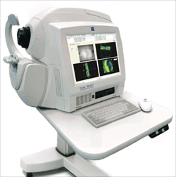 A noninvasive technique for imaging subsurface tissue structure with micrometer-scale resolution, the OPTICAL COHERENCE TOMOGRAPHY (OCT) is a relatively new technique for two and three-dimensional imaging of tissues at the histological level.
The technique is based on optical technology and commercially available fiber-optic components adapted for ophthalmic use. OCT is a non-invasive technique that does not utilize ionizing radiation to provide in vivo images. OCT has numerous potential clinical applications and, in effect, creates “optical biopsies” of tissues. OCT may be able to contribute to early diagnosis of neurologic diseases like mulitple sclerosis and Alzheimer’s. We frequently use it to evaluate glaucoma progression, macular degeneration, diabetic eye disease, macular holes, tumors, and even fitting scleral contact lenses.
 This new technology combines retinal photography with digital imaging. This allows instant viewing of retinal images by both the doctor and the patient, without the use of dilation drops. These images serve a dual purpose: first for documenting any existing eye disease, and second for establishing baseline images to compare against any future possible changes.
This method of examining and documenting the retina is incredibly more detailed and accurate than the conventional drawing method still in use by clinics that choose not to offer this new technology. We believe baseline images will promote earlier diagnosis of many abnormal vision conditions, many of which can lead to permanent visual loss if not caught and treated in a timely manner.
 We have three different versions of this test that maps the sensitivity of central and peripheral vision. Each test has its advantages and your doctor will order the most appropriate analysis for your condition. A visual field can be used to detect glaucoma, stroke, brain tumors, vision blocked by droopy eyelids, and many other neurological disorders. The test usually takes 1-5 minutes per eye and results can be interpreted immediately following testing. We include a visual field as part of our standard exam for diabetics or those experiencing worsening headaches.
Does My Insurance Cover This Test?
Medical insurance companies will reimburse for some of these procedures only when there is an existing retinal disease which the photos are documenting. We offer digital retinal imaging as a powerful way of viewing your central retina without the need for dilation. When used as part of your comprehensive exam, our baseline screening imaging (when no disease is present) is $22.00. We are very excited about the results in this technology and highly recommend Digital Retinal Imaging as an optional addition to your exam.